Small patients, major illnesses, enhance MRI to help infants and young children clinical precise treatment
Author:Cancer Channel of the Medical Time:2022.08.02
*For medical professionals for reading reference

Big Coffee Lecture Hall-Symposium on Clinical and Image diagnosis and treatment of Cardiology
""
In order to strengthen the communication and collaboration between the Film Department and the Clinical Department, the China Health Promotion and Education Association specially initiated and hosted the "Examination of Precision-Adult-Image and Clinical Diagnosis and Treatment" projects to help promote the intersection and integration of various disciplines, enhance the disease from the disease from Precision diagnosis to accurate treatment full -process management. The "medical community" sorted out the wonderful content of this project, and reinstated the wonderful content of the meeting in the form of a series of reports to readers.
"
In this issue, the editors have compiled a discussion of a multidisciplinary joint consultation (MDT) discussion about a rare case of puzzle -like nipple tumors reported by doctors such as Luojiang Hospital of Southern Medical University and Ma Antong and other doctors. The important role played during the MDT process of difficult diseases.
There are huge masses in intracranial in children who are less than one year old, enhancing MRI help diagnosis!
Polar clue papilloma is relatively rare in intracranial intakes from the vein epithelial cells or the slow growth of the venom cells in the brain. Mainly occur in the side brain, the fourth brain room, and the bridge horn area. Some literature reports can also occur in the saddle area, brain stem, brain essence and spinal cord [3-5].
This case of context clip -shaped papilloma -like cases shared by doctors such as Luo Minzhong and Ma Antong showed us image detection, especially enhanced magnetic resonance imaging (MRI) testing s help.
Case
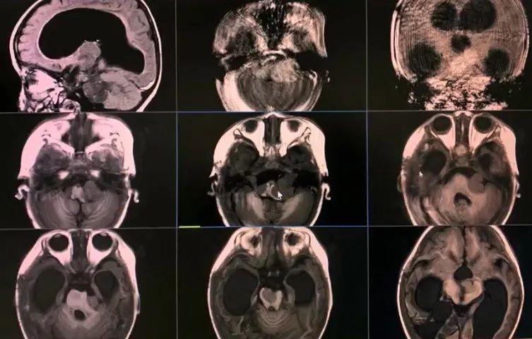
Men, in November, are normal for the birth and no special birth. Due to the "vomiting and difference for 1 week", he conducted a skull MRI inspection at the outer hospital, and found the right temporal top (7.5 mm × 9.5 mm × 6.5 mm), and then came to the hospital in May 2021. After admission, a huge tumor can be seen (Figure 1), which has accumulated multiple parts of the brain and caused the middle line structure to move left.
In this regard, Dr. Luo Kouyu of neurosurgery said that the recovery and prognosis of such children in clinical treatment and treatment have brought huge challenges to clinicians. The judgment of this issue requires the strong support of the video department.
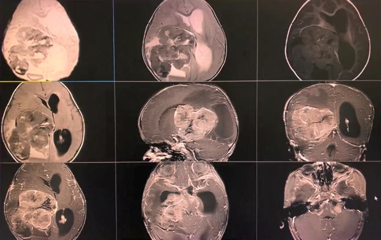
Figure 1 Children admitted to the hospital MRI detection image
Subsequently, Dr. Ma Antong of the Image Diagnosis Department introduced the video information of this case:
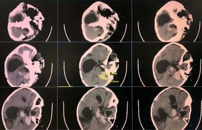
: The skull CT display:
The triangle of the triangle of the left side of the child is a leaf -shaped high -density lump with a clear border. The size is about 94 mm × 73 mm × 79 mm, and the CT value is about 50HU. The density is uniform. Multi -time -shaped low -density zone, the layered shadow is slightly high -density shadow behind the mass, considering it as local bleeding; Multi -nodule -shaped high -density shadows have clear boundaries.
像 像 像 ▎ MRI image display:
A huge leaf -shaped lump is seen in the triangle of the triangle of the side brain in the child. The border is clear. The T1 and T2 signals are mainly based on the T1 and T2 signals. The structure is about 19 mm to the left, and the stagnation of the stroke on the curtain is expanded; T2 signal, DWI sequence signal mixed, local high signal, ADC value is about 0.80mm2/s.
测 Enhance MRI monitoring display:
The intracranial lesions of the children are obviously imperious, and the triangle of the left cerebral ventricle triangle is cluster and the two -side bridge cerebelle corner area. The coronary image can be observed that the right side of the child's right side of the child is obviously oppressed, and the mid -line structure is severely shifted.
管 Magnetic resonance vascular imaging (MRA) display:
The lesion was supplied by the posterior cerebral arteries on the right side of the child, and the vascular surrounding blood vessels was stressed and transformed.
(Magnetic resonance Popp (H1-MRS) display:
The samples of the triangular lump area of the right side of the ventricle, the baseline of the spectrum is not stable, but you can see that the peak of the lipid (LIP) increase significantly (Figure 2).
Combined with various image detection results information, the masses of the triangular area of the left side of the child's left side of the ventricular chamber are considered as malignant tumor lesions, such as the vein plexus tumor, embryonic tumor, etc. The possibility of cerebrospinal fluid planting and metastasis in the nodule of the bridge horn area and the four -brain chamber side area.
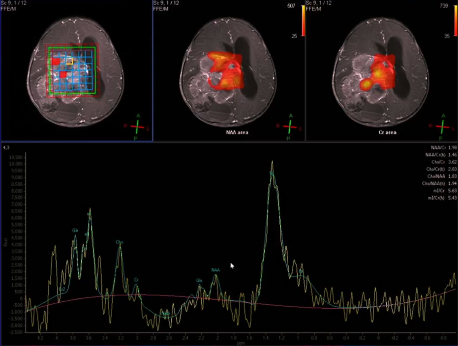
Figure 2 Children MRS test results
MRI helps clinicians to accurately track the condition of the children
Subsequently, Dr. Luo Kou added that the MRS of the children showed that its LIP peak increased significantly, the watches of the children's lesions were more malignant, and the necrotic tissue was more. After discussions by MDT, it was decided to provide laptopycin for children.
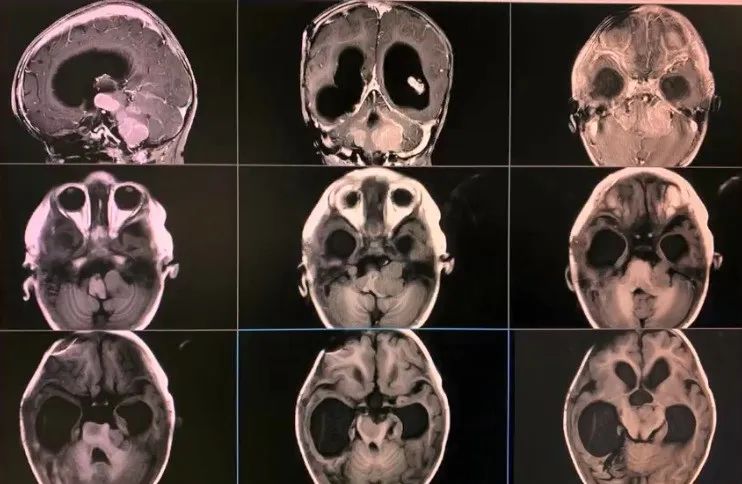
After the child's condition is stable, the drilling tumor cystic drainage+OMMAYA sac is enabled for the child's right pillow. The MRI prompts after the operation that the child's condition was stable, and the tumor did not see significantly (Figure 3).
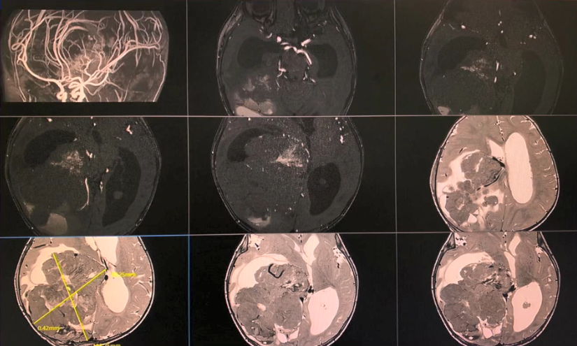
Figure 3 Children's right pillow drilling tumor cystic drainage+OMMAYA cysts after surgery review MRI results
As the child's condition gradually stabilizes, the family members of the child have stronger treatment confidence. After negotiating with the family members of the children, the family members were required to be strongly requested and prepared before surgery, and surgical resection for children.
All the lesions on the cranial scene during the operation were removed. The pathological test results of the postoperative showed that (the right temporal top pillow side ventricle) conforms to the vein clue papilloma (WHO III, Figure 4), which is consistent with pre -surgery image detection prediction results. In this regard, Director Guo Linlang of the Department of Pathology said that if the pathological test results such as the nuclear division elephant are alone, the tumor staging cannot be well judged, but the comprehensive judgment of the image detection results of the child can be given the above diagnosis.
Figure 4 Patients with lesions and pathological slices
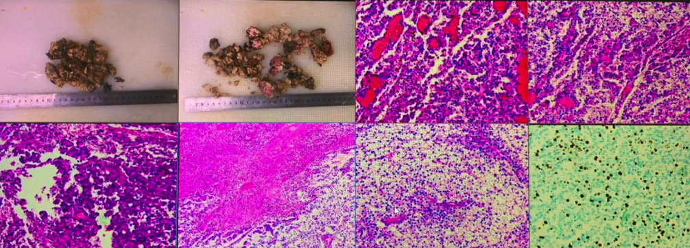
Unfortunately, the children have lung infections and abnormal liver function after surgery, and chemotherapy is not performed in time. At this time, the image MRI examination came in handy for postoperative condition monitoring.
Dr. Luo Kouyou introduced that the skull MRI was reviewed 40 days after surgery that the lesions under the intracranial scene of the child were significantly increased, and the children's brain stems were extremely severely oppressed (Figure 5).
Therefore, the children are immediately chemotherapy for the children. After 13 days of chemotherapy, the skull MRI shows that the patient's intracranial scene is still increasing. Fortunately, the surgical resection site has not recurred (Figure 6).
After the second stage of chemotherapy, the skull MRI testing prompts that the lesions of the children are controlled, the condition is stable (Figure 7), and further relief in subsequent treatment.
Fig
(Swipe left to view more)
Attach a wonderful case video
Summarize
The vein cluster papillary tumor has certain features in the MRI image, such as cauliflower or leaf -shaped mass in the brain, growing, and uneven edges. , T2WI signals are more mixed, DWI is equal or slightly lower signals, MRS is manifested as NAA, CR peaks, etc. [6].
MRI has a good soft tissue resolution, and can be multi -parameter, multi -sequence, and multi -functional imaging. For the relationships, boundaries, and surrounding structures in the lesion, it can be well displayed. Galex (钆 为 加) is a high concentration of a 1.0 mol/L comparison agent, while the concentration of other pyrine comparisons such as 钆teol and bispenamine is only 0.5 mol/L. It is suitable for examinations of patients with all -age parts of the whole body, and can also be used in full -moon newborns.
A study incorporated into 60 children under 2 years of age showed that after the child with a standard dose of the standard dose of the applied dosage enhanced examination, the image test results of children diagnosed 57 were consistent with the pathological test results. An adverse event related to the contrasting agent that occurs in the cymbal alcohol proves that the enhanced examination of the pyramidisol MRI can provide effective information on the diagnosis of various diseases of infants under 2 years of age, and at the same time have good safety and tolerance [7].
Precision image testing is one of the prerequisites for clinical "precise diagnosis to precise treatment". It can be described as the eyes of clinicians. In the future, it will also play a more important role in the full management of more patients.
Scan the code to watch
"Precise and Adverse" clinical and video combined with full case video
references:

[1] Koeller KK, Sandberg GD; Armed Forces Institute of Pathology. From the archives of the AFIP. Cerebral intraventricular neoplasms: radiologic-pathologic correlation. Radiographics. 2002 Nov-Dec;22(6):1473-505.
[2] Shi YZ, Chen MZ, Huang W, Guo LL, Chen X, Kong D, Zhuang YY, Xu YM, Zhang RR, Bo GJ, Wang ZQ. Atypical choroid plexus papilloma: clinicopathological and neuroradiological features. Acta Radiol. 2017 AUG; 58 (8): 983-990. [3] xiao a, xu j, he x, you c. extraventricular choroid plexus papilloma in the brainsster. J neurosurg pediatr. 2013 SEP; 12 (3): 247-50.
[4] Kuo CH, Yen YS, Tu TH, Wu JC, Huang WC, Cheng H. Primary Choroid Plexus Papilloma over Sellar Region Mimicking with Craniopharyngioma: A Case Report and Literature Review. Cureus. 2018 Jun 20;10(6): E2849.
[5] Bian Lg, Sun QF, Wu HC, Jiang H, Sun Yh, Shen JK. Primary Choroid Plexus Papilloma in the Pituitary Fossa: Case Report and Literate Review. Acta Neurochir (Wien); 2011 APRE; 851-7.
[6] Shen Lirong, Xu Zhifeng, Si Jianrong. MR analysis and clinical significance of the papillary tumor in the brain indoor veins [J]. Journal of Mudanjiang Medical College, 2019,40 (03): 32-35.
[7] Bhargava R, NOGA M. Safety and Effical of Gadobutrol-ENHANCED MRI in Patients AGED Under 2 Years-A Single-Center, Observational Study. Magn Reson Insights. 2013; 6: 1-12.
*For medical and health professionals only to understand information. These information cannot replace professional medical guidance in any way, nor should they be regarded as diagnosis and treatment suggestions. If such information is used to understand the purpose other than information, the platform and the author shall not bear the relevant responsibilities.
-End-
Submission/reprint/business cooperation, please contact: [email protected]


- END -
Thanks to the doctor for the 12 -year "companion" diagnosis and treatment, the guy went to the hospital to report to the hospital after the college entrance examination
Wuhan Evening News July 18th from the first consultation from 12 years ago, he has...
The fifth phase of prenatal ultrasound diagnosis standardized technology training course and the second meeting of the second meeting of the Hunan Obstetrics and Gynecology Ultrasound was successfully held

Huasheng Online News (Correspondent Long Xia Xiao Lihong) On July 9th, the fifth p...